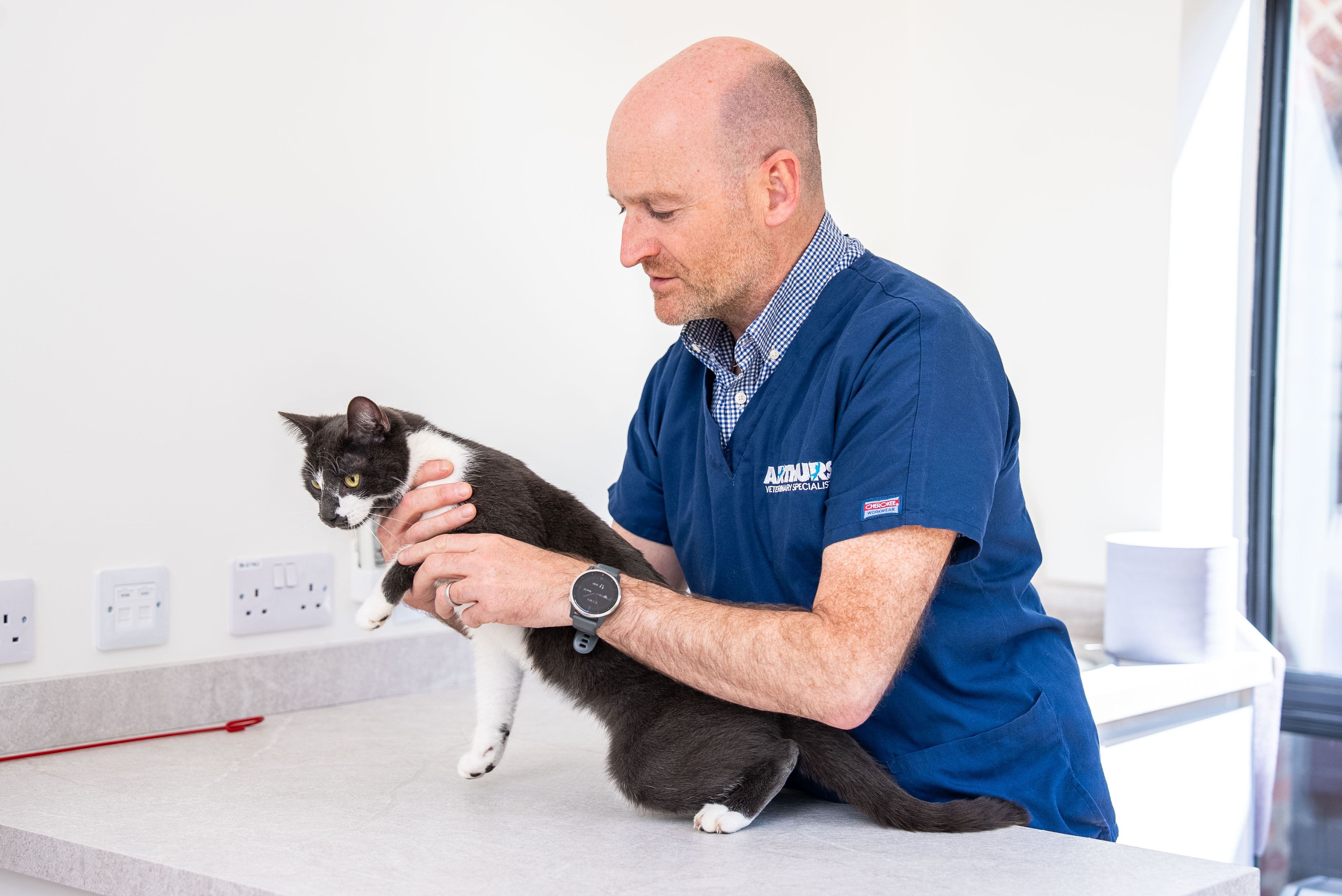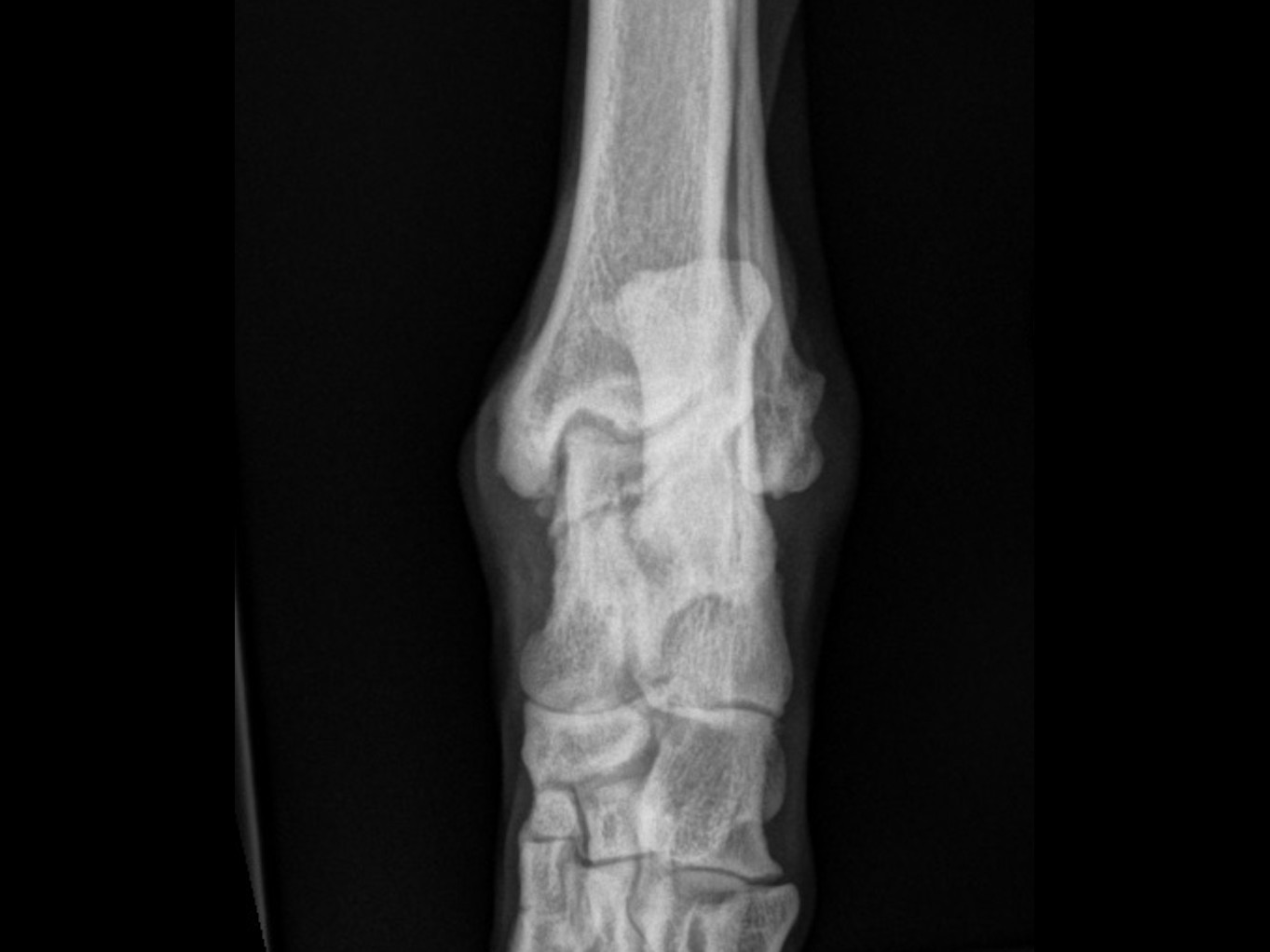
Shoulder osteochondrosis is an uncommon developmental condition of medium and large breed dogs where cartilage and bone on the humeral head fail to develop normally, causing forelimb lameness. Surgery to remove the loose flap gives excellent results in most dogs.
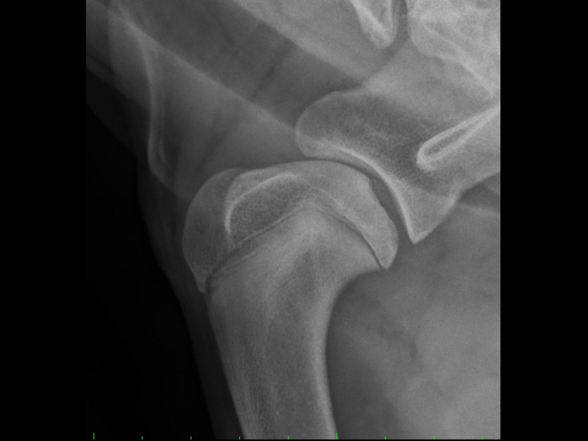
Shoulder osteochondrosis and secondary osteoarthritis is a condition that uncommonly affects medium and large breed dogs. The condition has a hereditary component, but inheritance is not simple and the condition is otherwise poorly understood. It is a condition whereby an area of cartilage and underlying bone on the back of the humeral head (the “ball” of the “ball and socket” shoulder joint) do not develop normally. The abnormal tissue detaches from the underlying bone, creating a loose flap which causes pain and inflammation.
It is typically diagnosed in young dogs around 6 to 10 months of age; signs include forelimb lameness and stiffness. Some dogs are affected severely, others only very mildly. Diagnostic tests include X-rays or CT scan; CT scan is much more accurate and gives much more information, so is preferred.
Management options depend on the severity of the condition:
For dogs that do not respond well, other options include implants to fill the defect in the humeral head — though these are not easy to place, are expensive, carry various possible complications, and the evidence supporting their use is limited. For dogs that respond poorly to all other options, arthrodesis (fusion) of the shoulder joint is performed; this is a complex procedure so is performed only rarely in dogs that are otherwise not coping.
Most dogs respond extremely well to surgery to remove the flap, with rapid resolution of the lameness and only minimal problems of minor stiffness and lameness in the future. It is important to understand that surgery is not a complete cure, as the underlying lesion / defect in the humeral head is not addressed and remains untreated. In the vast majority of dogs this has minimal consequence, but in a minority a residual or more obvious lameness is apparent.
Dogs that undergo a successful shoulder fusion (arthrodesis) do very well and are minimally to not lame.
Clear recovery guidance so you know what to expect at every stage.
Restricted activity and rest. Pain management and wound care. Short lead walks for toileting only. Your pet will have a follow-up check at 2 weeks for suture removal.
Gradually increasing lead walks. Continued restricted activity at home. Physiotherapy exercises as advised by our team to support healing and muscle rebuilding.
Follow-up imaging to check bone healing. Gradual increase in exercise duration and intensity based on progress. Most dogs show significant improvement by this stage.
Return to controlled normal activity, case dependent. Off-lead exercise can usually be gradually reintroduced. Full recovery is typically achieved within 3–4 months.
Answers to common questions about this procedure.
Whether you're a vet looking to refer or a pet owner wanting to learn more, we'd love to hear from you.
Refer a Case →