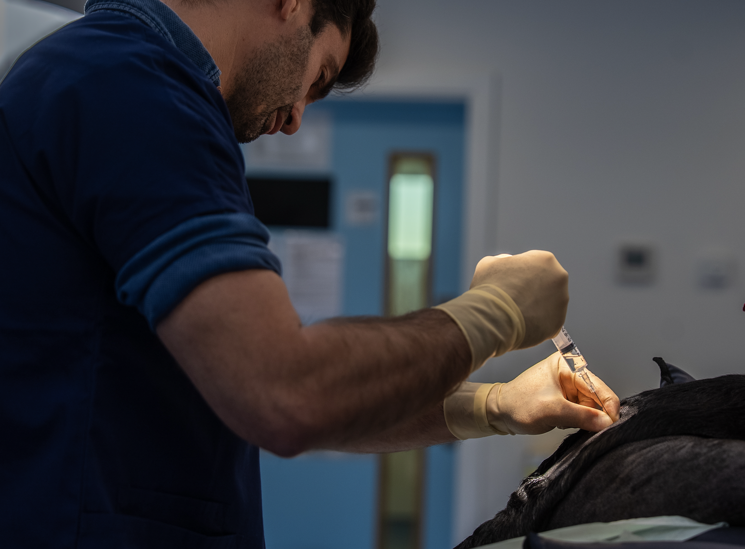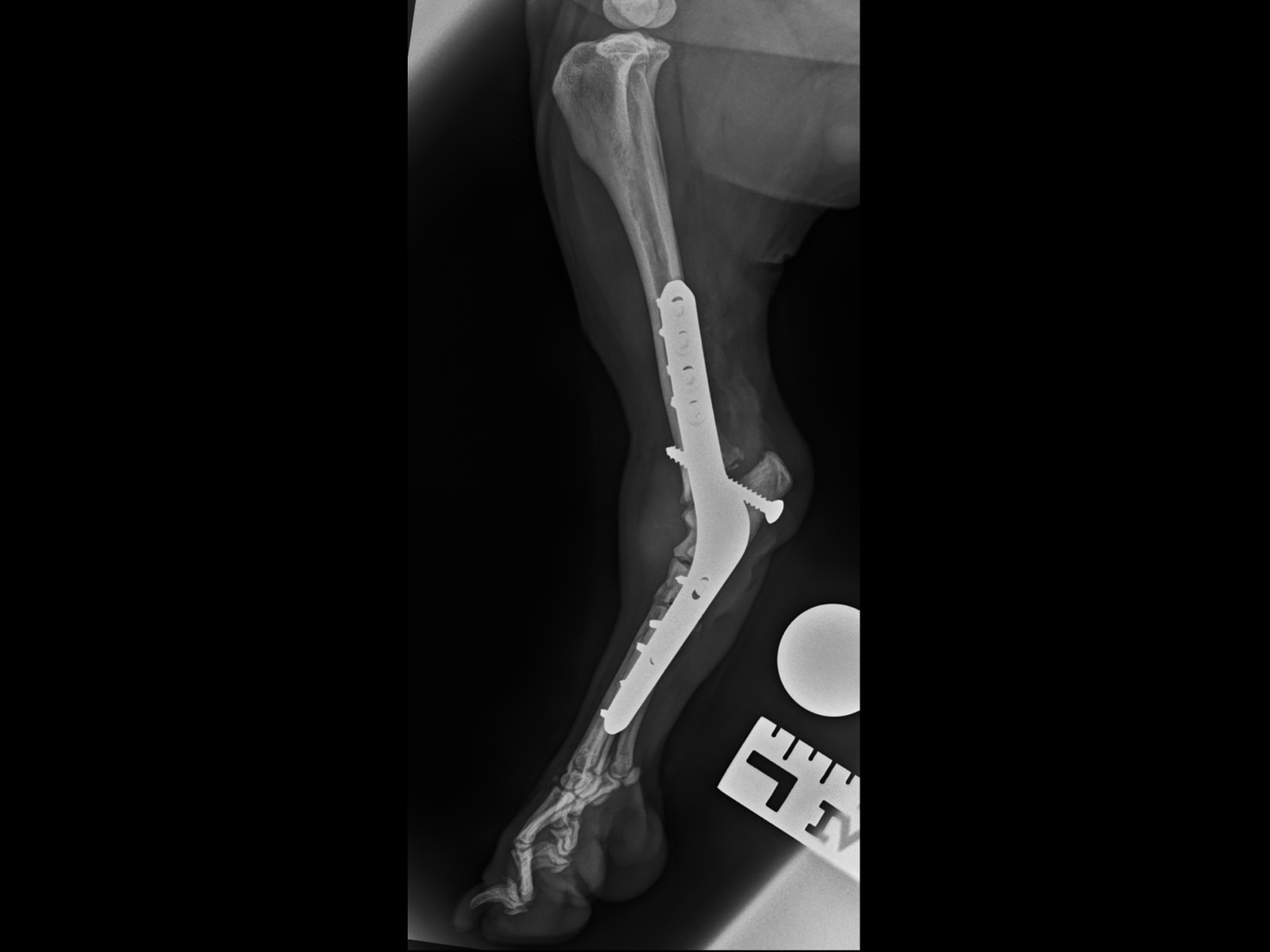
Hock osteochondrosis (OCD) is a hereditary developmental condition that uncommonly affects the hock (ankle) of medium and large breed dogs, where cartilage and bone of the talus fails to develop properly, causing lameness and arthritis.
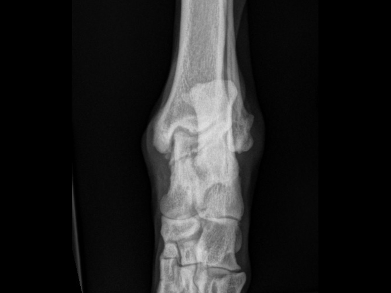
Hock Osteochondrosis (Osteochondritis Dissecans, OCD) is a hereditary developmental condition that uncommonly affects the hock (ankle joint) of medium and large breed dogs, including Labradors and Rottweilers.
One bone of the ankle joint is called the talus. It has two ridges — the medial and lateral trochlear ridges. In osteochondrosis, the bone and cartilage that form one ridge (usually the medial ridge) do not develop properly and fragment / fracture off. This means there is a loose portion of bone and cartilage within the ankle joint, and part of the weight-bearing surface is abnormal, causing joint inflammation, instability, swelling, thickening, arthritis (degenerative joint disease), stiffness and lameness.
It is a developmental disease, with the problem typically occurring between 3 and 6 months of age; however, in some dogs if the problem is mild, it may not be picked up until later in life. Diagnosis has three complementary parts: orthopaedic (physical) examination of the ankle joint, which is highly suggestive but not absolute; X-rays, which are good for diagnosis and for planning ankle replacement or fusion surgery; and CT scan, which is the gold standard, most accurate way to make the diagnosis and is particularly helpful if the lesion is subtle or if considering fragment removal.
Management options range from conservative care to salvage surgery:
Many dogs with hock osteochondrosis can be managed successfully with conservative care life-long. Where surgery is needed, outcomes vary by procedure.
Fragment removal has an unpredictable success rate — at best, 50% of dogs would see some improvement, but not a cure. Pantarsal arthrodesis (ankle fusion) provides excellent functional recovery with usually near normal hindlimb function, although the surgical procedure comes with a long list of potential complications, the most severe of which would be surgical site or implant infection, wound breakdown or skin necrosis (death). The published complication rate for arthrodesis is 30%, but we would estimate ours to be much lower at 10% or less.
Clear recovery guidance so you know what to expect at every stage.
Restricted activity and rest. Pain management and wound care. Short lead walks for toileting only. Your pet will have a follow-up check at 2 weeks for suture removal.
Gradually increasing lead walks. Continued restricted activity at home. Physiotherapy exercises as advised by our team to support healing and muscle rebuilding.
Follow-up imaging to check bone healing. Gradual increase in exercise duration and intensity based on progress. Most dogs show significant improvement by this stage.
Return to controlled normal activity, case dependent. Off-lead exercise can usually be gradually reintroduced. Full recovery is typically achieved within 3–4 months.
Answers to common questions about this procedure.
Whether you're a vet looking to refer or a pet owner wanting to learn more, we'd love to hear from you.
Refer a Case →