Advanced Diagnostics

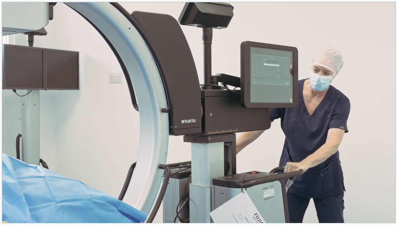
In-house CT scanning and digital X-rays so we can diagnose accurately and plan the best course of treatment for your pet.
Advanced Diagnostics

What Is This Procedure?

Our in-house diagnostic facilities allow us to investigate your pet's condition thoroughly and plan the most effective treatment. Having advanced imaging on site means we can often diagnose and plan surgery during a single visit, reducing the need for multiple appointments and anaesthetics.

Our diagnostic capabilities include:

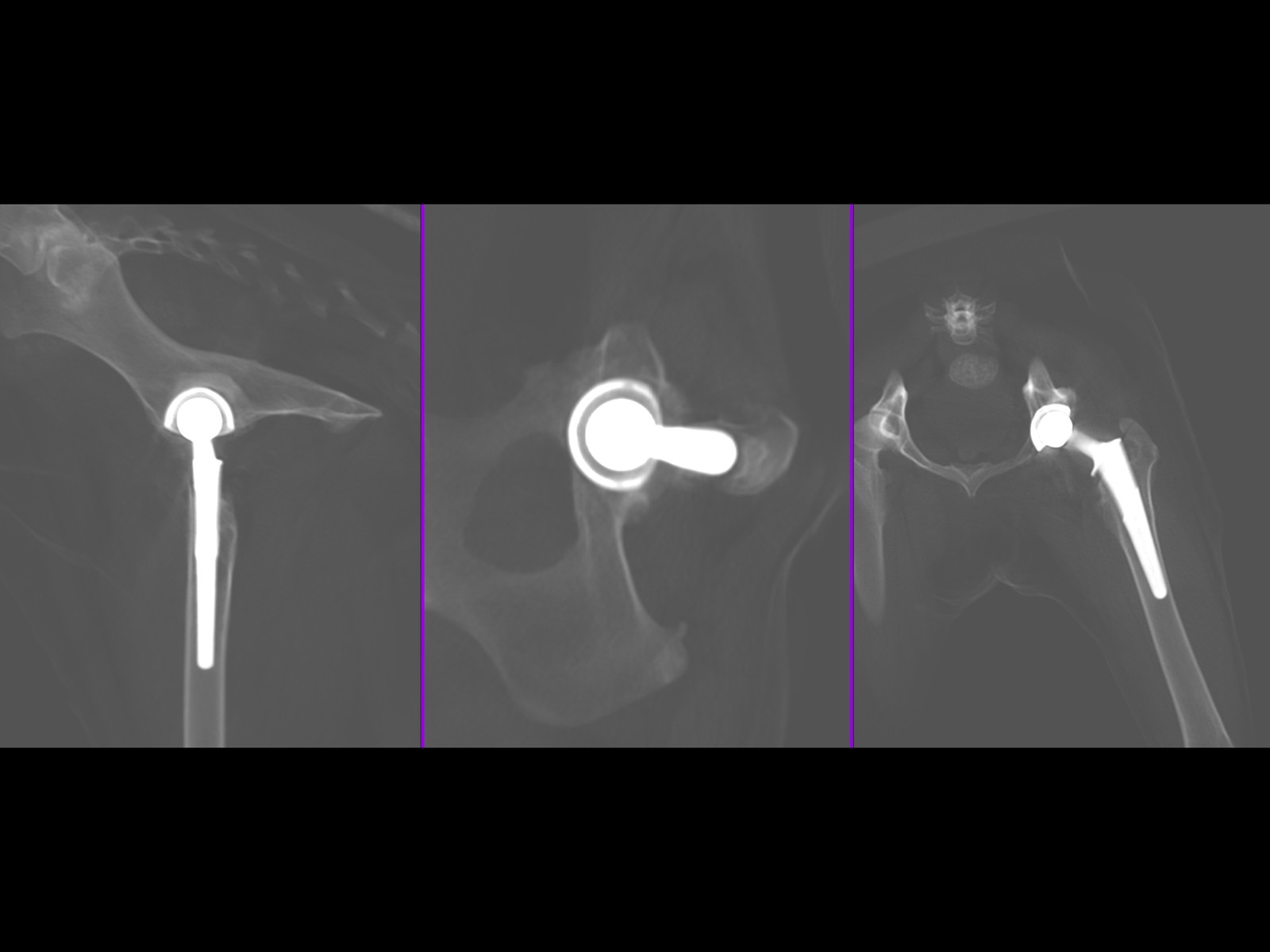
  • CT scanning — our 80/160 slice Canon CT scanner provides highly detailed 3D imaging of bones and joints, essential for planning complex orthopaedic surgery, identifying subtle fractures, and assessing conditions like elbow dysplasia and angular limb deformities. CT scans are also available as an outpatient service for referring practices.
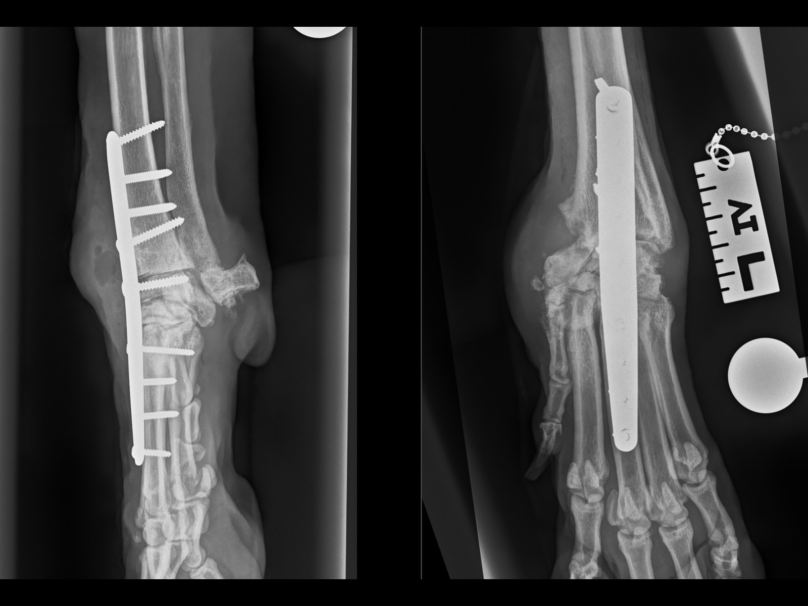
  • Digital radiography — high-quality digital X-rays provide rapid results for assessing fractures, joint conditions, and monitoring post-operative healing.
  • On-site laboratory — blood samples can be processed in-house for rapid results, including full biochemistry, haematology and electrolyte panels.

When Is It Recommended?

Advanced diagnostics are used throughout the referral process:

  • Initial investigation — when a pet is referred with lameness or an orthopaedic concern, imaging helps us reach an accurate diagnosis and understand the full extent of the problem
  • Pre-surgical planning — CT scanning is particularly valuable for planning complex procedures such as angular limb deformity corrections, where 3D printed bone models and surgical guides can be produced from the scan data
  • Monitoring recovery — post-operative X-rays confirm that fractures are healing and implants remain in position

Expected Outcomes

Advanced diagnostic imaging provides:

  • Accurate, definitive diagnosis to guide treatment decisions
  • Detailed pre-operative planning that improves surgical outcomes
  • Reduced need for exploratory surgery through non-invasive investigation
  • Clear visual evidence that can be shared with you and your referring vet
  • Rapid results, often within the same appointment

Recovery Timeline

Clear recovery guidance so you know what to expect at every stage.

1
0 – 2 WEEKS

Initial Recovery

Restricted activity and rest. Pain management and wound care. Short lead walks for toileting only. Your pet will have a follow-up check at 2 weeks for suture removal.

2
2 – 6 WEEKS

Controlled Recovery

Gradually increasing lead walks. Continued restricted activity at home. Physiotherapy exercises as advised by our team to support healing and muscle rebuilding.

3
6 – 8 WEEKS

Follow-Up Assessment

Follow-up imaging to check bone healing. Gradual increase in exercise duration and intensity based on progress. Most dogs show significant improvement by this stage.

4
10 – 12 WEEKS

Return to Normal Activity

Return to controlled normal activity, case dependent. Off-lead exercise can usually be gradually reintroduced. Full recovery is typically achieved within 3–4 months.

Frequently Asked Questions

Answers to common questions about this procedure.

Do you share the images with my own vet?

Always. We send the full images and a written report to your referring vet so they have everything they need for your pet's ongoing care.

Is advanced imaging safe for my pet?

Yes. Modern imaging is low-risk and our experienced clinical team monitor your pet closely throughout. Where sedation is needed it is short-acting and carefully tailored.

What is advanced diagnostic imaging used for?

Advanced diagnostics — including CT, ultrasound and high-detail X-ray — let us see exactly what is happening inside the joint or bone, so we can plan the best treatment with confidence.

Related Services

GET STARTED

Ready to Refer a Patient?

Whether you're a vet looking to refer or a pet owner wanting to learn more, we'd love to hear from you.

Refer a Case →