Elbow Dysplasia

Elbow dysplasia and secondary arthritis is a hereditary condition affecting many medium and large breed dogs. It is a spectrum of diseases; management ranges from conservative care to arthroscopic and salvage surgery.

Elbow Dysplasia

What Is This Procedure?

Elbow dysplasia and secondary arthritis is a condition that affects many medium and large breed dogs. It is a hereditary condition, but inheritance is not simple and the condition is otherwise poorly understood. Elbow dysplasia is a spectrum of diseases including:

  • Elbow incongruency (mis-shapen joint) — believed to contribute to elbow dysplasia in all dogs, but difficult to define except in the most obvious cases.
  • Medial Coronoid Process disease / fragmentation — the most common form. The medial coronoid process is a part of the ulna bone that makes part of the elbow joint; it can be fragmented, abnormal in shape or abnormal in texture. CT scan is about 80% sensitive to fragmentation.
  • Arthritis / degenerative joint disease — happens in almost all cases of elbow dysplasia, including cartilage erosion through the medial compartment of the elbow joint, ranging from very mild to total erosion.
  • Kissing lesion — a focal area of cartilage erosion of the medial humeral condyle, present in most dogs with elbow dysplasia to a greater or lesser extent.
  • Osteochondrosis of the medial humeral condyle — a focal defect of cartilage and underlying bone; quite uncommon, which is fortunate as there is no good treatment.
  • Ununited Anconeal Process — typically affects large breed dogs and German Shepherds; when caught early (less than 12–18 months of age) the fragment can be screwed / pinned back into position, often combined with a releasing cut in the ulna bone (ulna osteotomy).

The most sensitive tests for elbow dysplasia are CT scan and arthroscopy (keyhole surgery); X-rays can be used but are not very sensitive.

When Is It Recommended?

Management options depend on the severity and stage of the disease:

  • Conservative (non-surgical) management — restricting exercise to a level that does not exacerbate lameness, avoiding boisterous / high-impact activity, short frequent walks, keeping body condition slim, and using anti-inflammatory pain-killers and/or joint supplements as necessary. Normally recommended for dogs with relatively mild lameness or mild CT changes.
  • Arthroscopic surgery of the elbow — to inspect and remove an abnormal medial coronoid process and document cartilage damage; sometimes combined with ulna osteotomy. Normally recommended for dogs with definite fragmentation, more than mild lameness, that have not responded to conservative management.
  • Elbow injections — steroids (short-term relief but likely more cartilage damage), viscoelastic lubricants (limited evidence), platelet rich plasma (limited evidence), and stem cell injections harvested from a fat graft (a novel treatment with some evidence it could be effective).
  • Other surgery options — ulnar osteotomy, Sliding Humeral Osteotomy (SHO), and Canine Unicompartmental Elbow arthroplasty (CUE); these have unproven and variable outcomes, are quite invasive, and some have relatively high complication rates and are expensive.
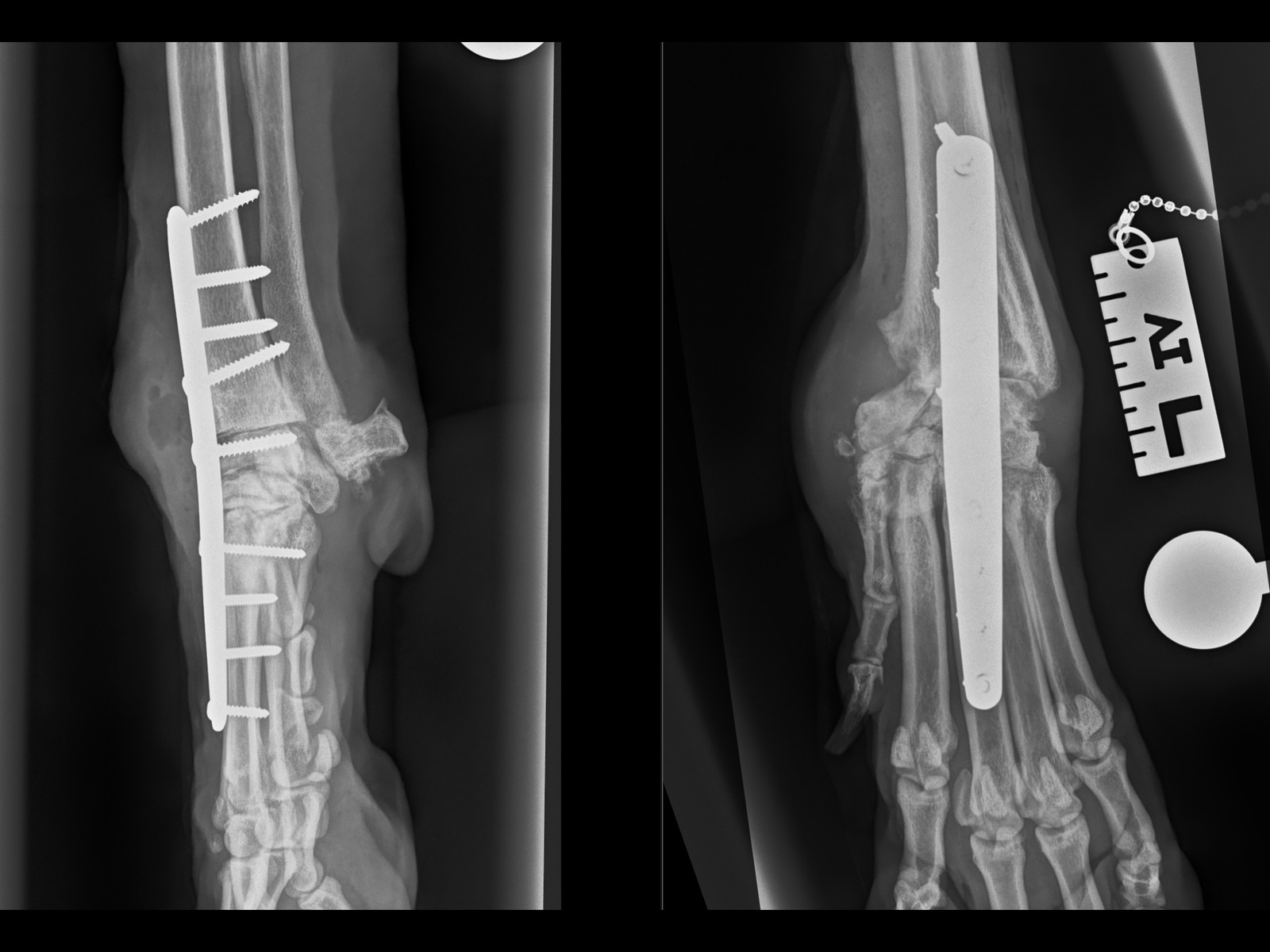
  • Salvage surgery — offered when everything else has failed and the dog is really struggling: Total Elbow Replacement, elbow arthrodesis (fusion), or amputation. Very few dogs get so bad as to consider these options.

Expected Outcomes

It is important to understand that surgery is not curative and lameness / arthritis will progress regardless of surgery. In younger dogs (puppies and young adults), it is thought that about 60–70% of dogs improve (but are not cured) following arthroscopic surgery, and that this improvement lasts for months to a few years. However, 30–40% of dogs do not improve following arthroscopic surgery, and surgery can make the lameness worse, although this is very rare. In middle / older age dogs, these odds decrease to 50/50 at best.

Recovery Timeline

Clear recovery guidance so you know what to expect at every stage.

1
0 – 2 WEEKS

Initial Recovery

Restricted activity and rest. Pain management and wound care. Short lead walks for toileting only. Your pet will have a follow-up check at 2 weeks for suture removal.

2
2 – 6 WEEKS

Controlled Recovery

Gradually increasing lead walks. Continued restricted activity at home. Physiotherapy exercises as advised by our team to support healing and muscle rebuilding.

3
6 – 8 WEEKS

Follow-Up Assessment

Follow-up imaging to check bone healing. Gradual increase in exercise duration and intensity based on progress. Most dogs show significant improvement by this stage.

4
10 – 12 WEEKS

Return to Normal Activity

Return to controlled normal activity, case dependent. Off-lead exercise can usually be gradually reintroduced. Full recovery is typically achieved within 3–4 months.

Frequently Asked Questions

Answers to common questions about this procedure.

No items found.

Related Services

GET STARTED

Ready to Refer a Patient?

Whether you're a vet looking to refer or a pet owner wanting to learn more, we'd love to hear from you.

Refer a Case →