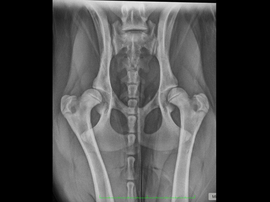
Hip luxation means a dislocated hip. This is usually caused by a traumatic event such as a fall, landing awkwardly, or being hit by a motor vehicle.
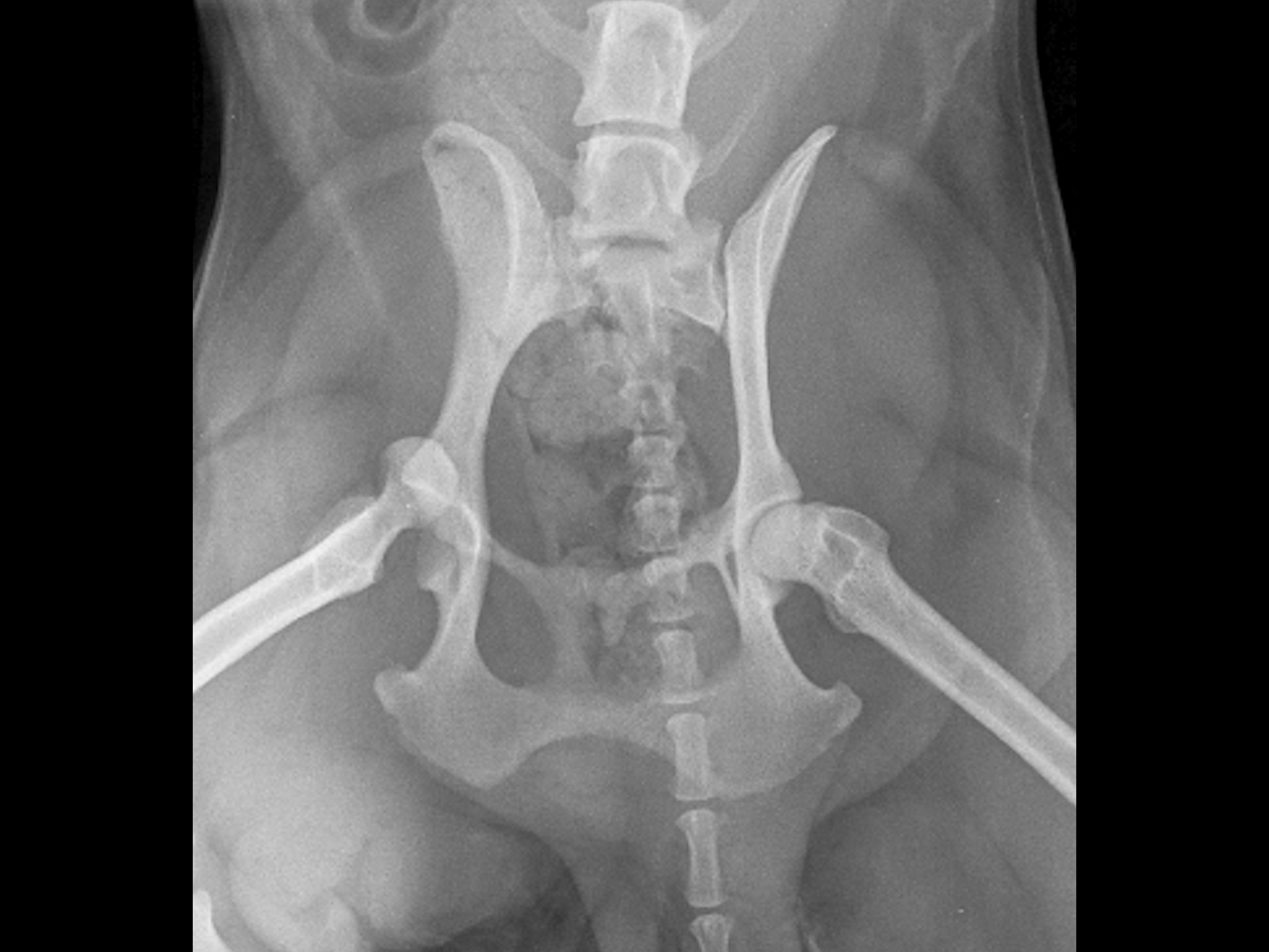
Hip luxation means a dislocated hip.
This is usually caused by a traumatic event such as a fall, landing awkwardly, or being hit by a motor vehicle.
Sometimes an underlying problem such as hip dysplasia can make a dog or cat more prone to hip dislocation.
Almost always, the signs of hip luxation are sudden onset severe to non-weight bearing lameness of a hindlimb; usually caused by a traumatic event.
If we are suspicious that your pet is suffering from a luxated hip, investigation involves:
If your pet is diagnosed with a dislocated hip, a variety of treatment options are available; but the right choice for you and your pet depends on a number of factors, which we will discuss, and recommend the best option for your pet.
Treatment options include:
Expected outcome after surgical management should be excellent, with a normal, pain-free range of joint movement and function expected. Most dogs will use the operated leg within a few days of surgery; have a mild to moderate lameness by 2 months post-op and be free of lameness and exercising normally by 4 month post-op.
Dogs or cats recovering from this condition should have significant improvement in limb function and able to comfortably walk, sit, climbing stairs, stand, run, getting into a car, play and exercise; normal to near-normal function can be expected.
Clear recovery guidance so you know what to expect at every stage.
Restricted activity and rest. Pain management and wound care. Short lead walks for toileting only. Your pet will have a follow-up check at 2 weeks for suture removal.
Gradually increasing lead walks. Continued restricted activity at home. Physiotherapy exercises as advised by our team to support healing and muscle rebuilding.
Follow-up imaging to check bone healing. Gradual increase in exercise duration and intensity based on progress. Most dogs show significant improvement by this stage.
Return to controlled normal activity, case dependent. Off-lead exercise can usually be gradually reintroduced. Full recovery is typically achieved within 3–4 months.
Answers to common questions about this procedure.
Whether you're a vet looking to refer or a pet owner wanting to learn more, we'd love to hear from you.
Refer a Case →