Hip dysplasia is a common orthopaedic problem of dogs, especially medium to giant breeds, caused by abnormal development of the hip joint in the puppy. Both surgical and non-surgical treatments are available.
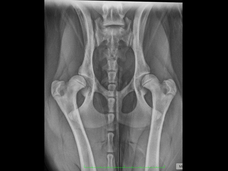
Hip dysplasia is a common orthopaedic problem of dogs, especially medium to giant breeds. It occurs due to abnormal development and growth of the hip joint in the puppy. Both hips are usually affected, but only one side may show symptoms, and the onset of clinical signs (pain or lameness) may not occur on both sides at the same time.
Hip dysplasia is manifest early in life by varying degrees of laxity of the muscles and ligaments around the hip joint, which results in instability and malformation of the joint components. Arthritis is the long-term consequence of undetected or untreated hip dysplasia, due to the wear and tear which affects the joint surfaces as a result of the laxity.
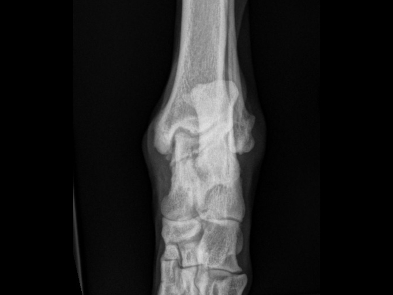
The clinical signs of hip dysplasia are hind limb lameness, hind limb stiffness, bunny hopping, reluctance to lie down, sit, or get up, reluctance to go upstairs or jump onto things (e.g. into the car boot), and tiring on a walk. These signs are seen in puppies from around 6 to 18 months of age, or adult dogs 3–6 years of age. Investigation involves clinical examination, followed by sedation for hip X-rays or CT scan, and hip manipulation to test the degree of laxity and conformation.
Several treatment options are available, both surgical and non-surgical:
Prognosis with non-surgical management is good in many cases — up to 70% of puppies with hip dysplasia respond to conservative management and are never bad enough to need surgery even when they are adults. It is widely accepted that in dogs with hip dysplasia, arthritis progresses more quickly in overweight dogs.
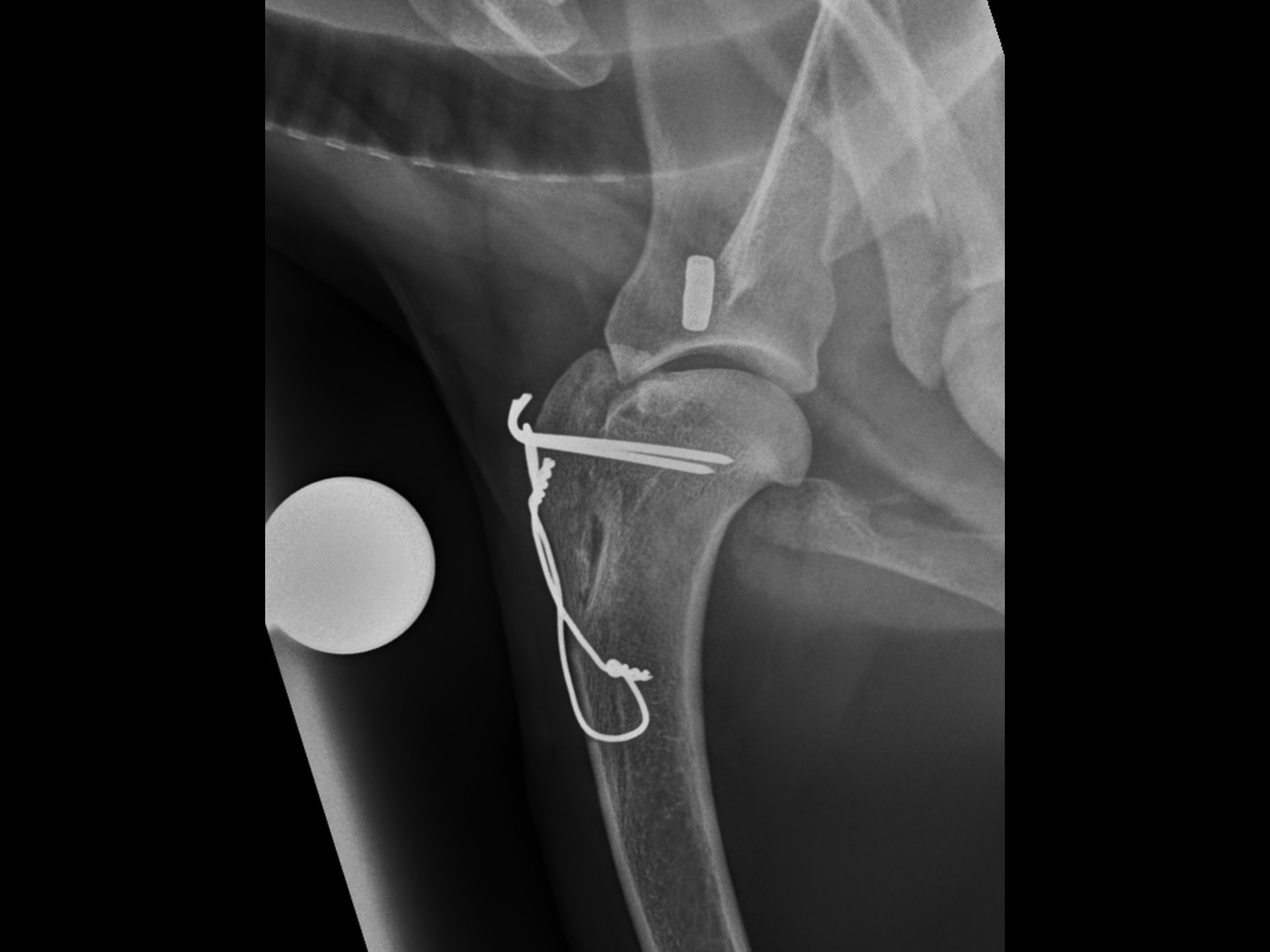
Where surgery is needed, Total Hip Replacement is the gold standard for regaining as near normal hip function as possible and produces excellent results for pain-free hip function. Deciding which treatment is best is not always easy and requires careful assessment and discussion on a case-by-case basis.
Clear recovery guidance so you know what to expect at every stage.
Restricted activity and rest. Pain management and wound care. Short lead walks for toileting only. Your pet will have a follow-up check at 2 weeks for suture removal.
Gradually increasing lead walks. Continued restricted activity at home. Physiotherapy exercises as advised by our team to support healing and muscle rebuilding.
Follow-up imaging to check bone healing. Gradual increase in exercise duration and intensity based on progress. Most dogs show significant improvement by this stage.
Return to controlled normal activity, case dependent. Off-lead exercise can usually be gradually reintroduced. Full recovery is typically achieved within 3–4 months.
Answers to common questions about this procedure.
Whether you're a vet looking to refer or a pet owner wanting to learn more, we'd love to hear from you.
Refer a Case →